Intraocular tumors
What you need to know about intraocular tumors


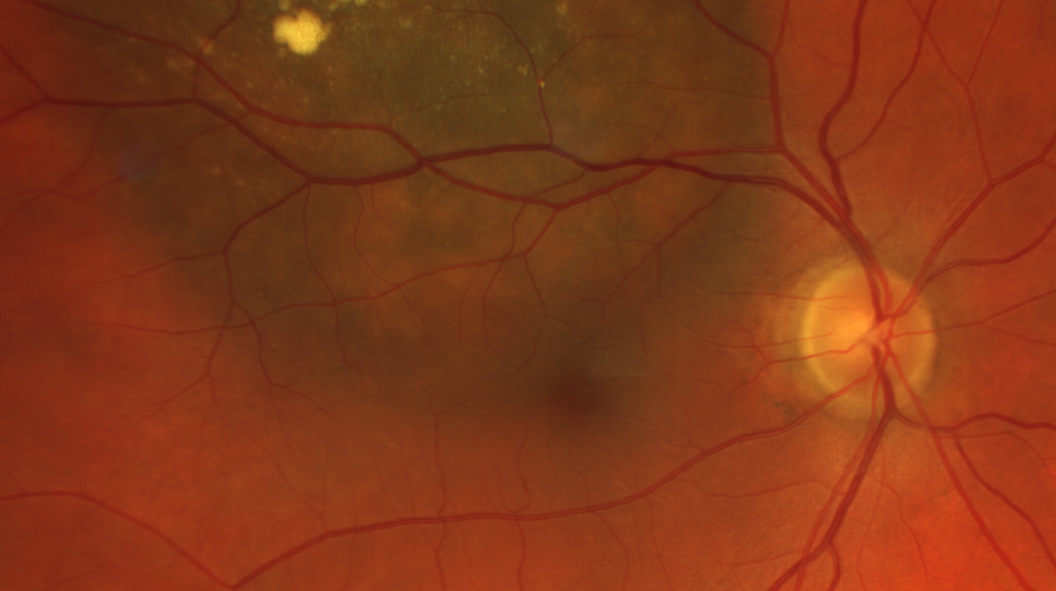
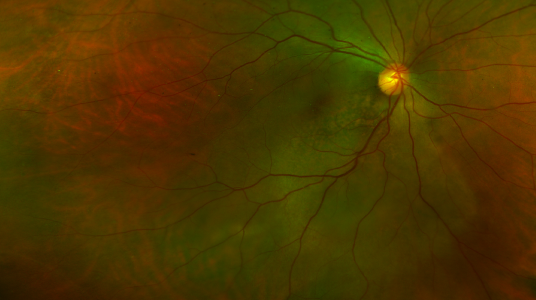
Anatomy
The eyeball is made up of three tissue layers that form its outer structure, and transparent substances that fill the inside.The eye’s outer layers (the structure):
The inside of the eye (contents):
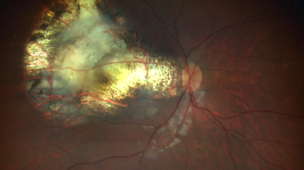
Uveal Melanoma
Uveal melanoma is the most common primary intraocular cancer in humans.
Intraocular lymphoma
Intraocular lymphoma is composed of several entities, depending on whether it involves uveal or retinal tissue.
Uveal Metastasis
Metastases from another cancer may appear in one or both eyes.

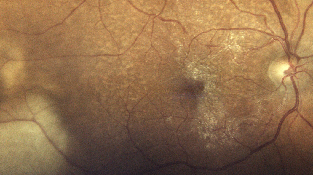
Choroidal hemangioma
Choroidal hemangiomas are benign intraocular lesions, whether circumscribed or diffuse.
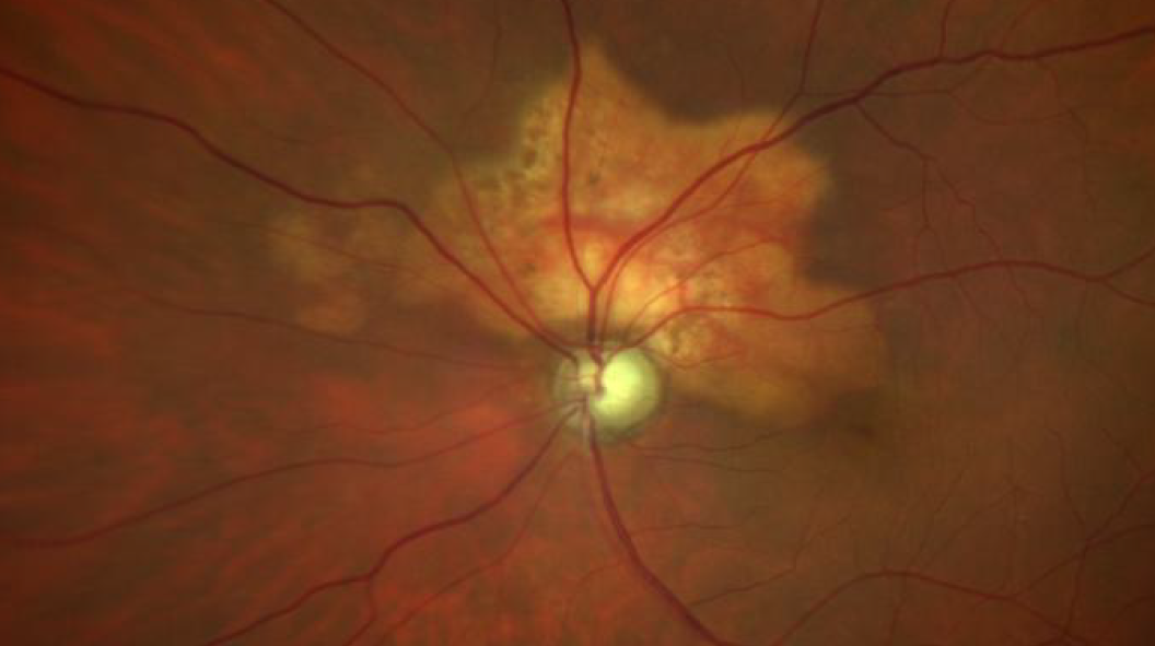
Retinoblastoma
Retinoblastoma is the most common intraocular cancer in children, although it is a rare cancer.
Other intraocular tumors
Other less common tumors can also develop inside the eye, whether cancerous or not.