Choroidal Hemangioma
Everything you need to know about choroidal hemangioma
Choroidal hemangioma is a benign vascular tumor of the choroid, composed of abnormal blood vessels, causing deformation of the choroid. It should not be confused with exudative chorioretinopathy secondary to thickening and tortuosity of choroidal vessels associated with the Sturge-Weber syndrome.
No risk factors have been identified to date. However, alterations of the GNAQ gene in the cells forming the wall of choroidal vessels have been described.
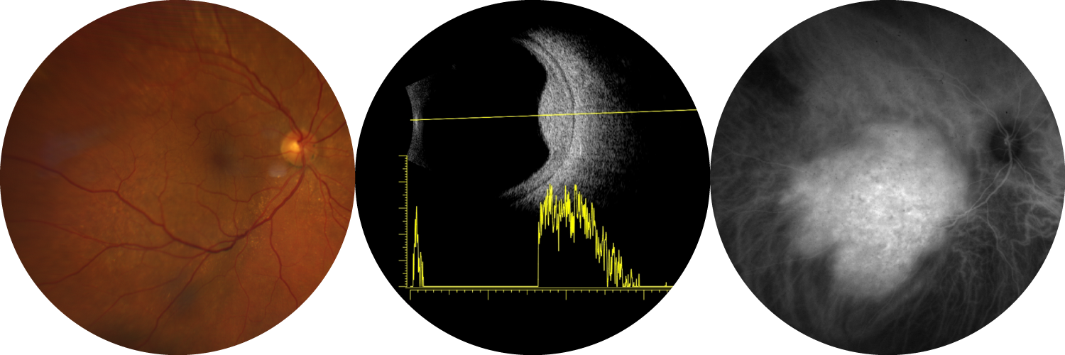
Due to its rarity, choroidal hemangioma is often difficult to diagnose. It may be discovered incidentally during retinal photography (orange-red lesion) and macular optical coherence tomography (OCT), by identifying a deformation of the choroid. More commonly, it can cause a decrease in visual acuity, a spot in the visual field (scotoma), and/or repetitive flashes (photopsias) caused by retinal detachment related to the choroidal hemangioma. Diagnosis relies on multimodal imaging, combining retinal photography, indocyanine green retinal angiography, and ocular ultrasound.
In the absence of decreased visual acuity caused by the hemangioma, no specific treatment is required. However, if vision is affected, treatment aims to dry out the lesion without destroying it. Treatment options include:
- administration of photodynamic therapy with verteporfin
- or radiotherapy, usually by proton therapy
Currently in France, due to limited availability of verteporfin, the first-line treatment for circumscribed hemangioma is proton therapy. The doses administered are about three times lower than those used for uveal melanoma, allowing preservation of retinal tissue.
From an ophthalmological standpoint, follow-up will be determined by the referring ocular oncologist, but it must be long-term since local exudative recurrences may occur, even more than 5 years after initial treatment. Proton therapy provides a more durable response than photodynamic therapy. Bilateral choroidal hemangiomas are extremely rare.
From a general standpoint, since this is a benign tumor, no specific systemic follow-up is required.