Intraocular Lymphomas
Everything you need to know about intraocular lymphomas
Intraocular lymphoma is a malignant tumor resulting from a monoclonal proliferation of immune system cells called lymphocytes. These are usually B lymphocytes that transform into lymphoma cells. It is always a non-Hodgkin lymphoma. In reality, there are two very different entities: vitreoretinal lymphoma and uveal lymphoma.
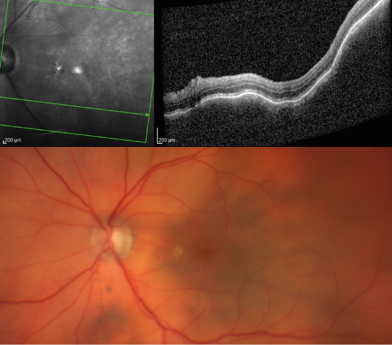
Vitreoretinal Lymphoma
Vitreoretinal lymphoma is usually a large B-cell lymphoma, meaning an aggressive lymphoma (high grade). It can be:
- a primary oculocerebral lymphoma, a cancer that develops initially in the eye and/or central nervous system. There is no risk of spreading outside the eye and central nervous system.
- a systemic lymphoma, a cancer that originates elsewhere in the body and spreads to the eye and/or central nervous system.
Uveal Lymphoma
Uveal lymphoma, usually located in the choroid, belongs to the group of ocular adnexal lymphomas (which can affect surrounding eye tissues such as the conjunctiva or the orbit). There are several subtypes of uveal lymphoma, most of which are low-grade indolent lymphomas, while a minority are more aggressive, disseminated, and considered high-grade lymphomas.


The risk factors for developing intraocular lymphoma depend on the subtype of lymphoma. In general, it is a disease of adults over the age of 50.
Clinical presentation will depend on the type of lymphoma (vitreoretinal or uveal), as well as on extraocular locations (particularly cerebral).
Vitreoretinal Lymphoma
It usually presents with vision loss, caused by vitreous opacities mimicking ocular inflammation (uveitis) and/or the appearance of white retinal lesions. Diagnosis relies on ophthalmic imaging (fundus photography, optical coherence tomography – OCT) and ophthalmic biological sampling. The level of biological markers (IL-6 and IL-10) is usually measured in the aqueous humor of the anterior chamber. In cases of a significant elevation of IL-10, a diagnostic vitrectomy is usually performed to confirm the diagnosis. Brain MRI, 18F-FDG PET scan, and cerebrospinal fluid sampling are typically part of the initial workup, whether the lymphoma is primary or not.
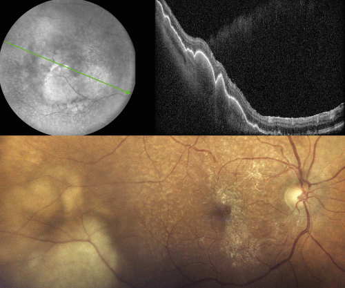
Uveal Lymphoma
It usually presents with vision loss due to choroidal deformation without specific pigmentation, and/or retinal detachment caused by the tumor. Diagnosis is based on ruling out other intraocular tumors (multimodal imaging) and performing a tumor biopsy.
Treatment depends on the type of lymphoma diagnosed, under the responsibility of a hemato-oncologist, or a neuro-oncologist in cases of oculocerebral lymphoma.
For vitreoretinal lymphoma, first-line treatment usually consists of high-dose systemic methotrexate. Management of this type of lymphoma requires a specialized opinion through a dedicated multidisciplinary tumor board of the LOC network, certified by the French National Cancer Institute.
For uveal lymphoma, treatment depends on several factors:
- whether the disease is limited to the eye or disseminated
- the patient’s age and life expectancy
- the patient’s comorbidities
- prognostic factors specific to each lymphoma subtype
Treatment may involve radiotherapy and/or immunochemotherapy, following multidisciplinary consultation in hemato-oncology.
Ophthalmologic follow-up will be specified by the referring ocular oncologist, depending on the treatments undertaken and their effectiveness.